Sperm usually reach the egg in the portion of the fallopian tube closest to the ovary within 90 minutes after ejaculation. Mitochondria (tiny energy sources for cells) in the midpiece of sperm cause the tail of the sperm to lash about. This flagellation, or lashing out, propels the sperm through the woman's vagina and into her tubes. Of the average 300 million sperm present in each ejaculation, an estimated 2,000 eventually reach the fallopian tube containing the ovum. Only 50 sperm may actually reach the egg. The remaining sperm are either killed by the acidic environment of the vagina or by entering the wrong fallopian tube. Only one sperm penetrates and fertilizes the ovum. The others surround the egg and secrete the enzyme hyaluronidase to soften the gelatinous covering of the egg, the zona pellucida. Once penetrated, the ovum's surrounding membrane thickens to prevent other sperm from entering.
Fertilization is completed when the nucleus of the head of the sperm fuses with the nucleus of the egg. At this time, the sex of the zygote is determined by the presence or absence of a Y chromosome.
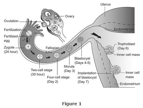
Within 24 to 36 hours, the single‐cell zygote begins dividing exponentially as it moves along the fallopian tube. One cell becomes two, two becomes four, four becomes eight, and so forth.
Implantation
Within a week after conception, the zygote becomes a blastocyst, or a hollow ball of about 100 cells, no larger than it was before cell division began. After floating in the uterus for about 3 days, the blastocyst attaches to the endometrium, or inner lining of the uterus. The outer cells of the blastocyst, or the trophoblasts (which form the trophectoderm), secrete enzymes that dissolve layers of uterine lining, allowing the blastocyst to firmly attach to the endometrium. This implantation occurs about a week after conception. After implantation, and for the first 8 weeks of gestation, the zygote is referred to as an embryo. (See Figure for illustration of the early development of the embryo.) Following the first 8 weeks until birth, it is referred to as a fetus.
During implantation, the rate of cell division increases, and the first signs of specialized organs and tissues appear. For example, a small indentation, the neural groove, forms and later develops into the fetus's brain and nervous system. After implantation, other cells, including the trophoblasts, develop into the fetus's placenta, umbilical cord, and amniotic sac.
The first trimester
The placenta is a disk‐shaped structure of tissue that forms along the uterine wall on one side and attaches to the fetus via the umbilical cord. The placenta's function is to pass oxygen, nourishment, and antibodies from the mother's blood to the developing fetus. Similarly, waste products from the fetus are passed to the mother for elimination. Early in gestation the placenta begins secreting the hormone human chorionic gonadotropin ( HCG). HCG inhibits menstrual periods by preserving the corpus luteum (the empty Graafian follicle which releases the mature ovum) during the early stages of pregnancy. The hormone is present in a woman's blood and urine soon after conception and is the basis of most common pregnancy tests: If HCG is present, the test is positive, and the woman is pregnant. Home‐based tests are more likely than laboratory tests to give false negative results, which may lead the woman to think she is not pregnant when she is. Thus, home pregnancy tests are not as reliable as laboratory tests.
Formed during the fifth week of embryonic development, the umbilical cord carries blood to and from the fetus via one vein and two arteries. Fetal blood circulates through the chorionic villi, which are small fingerlike projections in the placenta. The mother's and infant's circulatory systems remain totally separate, yet oxygen, carbon dioxide, waste products, nutrients, viruses, and assorted drugs can cross the membrane of the chorionic villi.
After the first trimester, the placenta also secretes large amounts of progesterone and estrogen. Many of the physical symptoms of pregnancy can be traced to the actions of these two hormones. Estrogen and progesterone stimulate enlargement of the reproductive organs and relaxation of associated ligaments, stimulate development of the uterine lining and mammary glands, and prevent contractions of the uterus. Another hormone produced by the placenta, placental lactogen, prepares the mammary glands to secrete milk.
Two membranes surround the embryo; the inner membrane is known as the amnion, and the outer membrane is called the chorion. The fetus floats in the waterlike amniotic fluid that fills the amniotic sac (formed from the fusion of the amnion and chorion). Amniotic fluid cushions the developing fetus against injury and shock and provides constant temperature in the amniotic sac.
When a woman suspects that she is pregnant, she should have her status confirmed as soon as possible. One method is to administer an HCG‐based pregnancy test. Another is ballottement, a type of pelvic examination in which a physician or nurse feels for a fetus in the uterus. Pregnancy may also be verified when a doctor hears a fetal rushing sound, or uterine souffle, by listening through a stethoscope placed on the women's abdomen.
Fetal development occurs in cephalocaudal order, beginning with the head and ending with the lower body and extremities. This sequence of development results in the head of a typical fetus being disproportionately larger than the rest of its body.
Most of the fetus's systems and structures begin to form during the first 12 weeks of gestation. Three layers of cells differentiate to become the various body organs. The ectoderm (outermost layer) forms the sensory organs, skin, and nervous system. The mesoderm (middle layer) forms the connective tissues, muscles, skeleton, and circulatory and reproductive systems. The endoderm (innermost layer) forms the digestive, respiratory, and glandular systems.
The digestive and respiratory organs begin limited functioning by about week 7. The gonads have also already begun developing, even though fetus's gender is not yet externally visible.
The embryo weighs approximately 1/30 of an ounce and is 1 1/4 inches long around week 8. The fetus's tongue, lips, ears, nose, and eyes can be seen. The fetus's head is much larger than the rest of its body due to rapid growth of the brain. Arms, hands, legs, feet, and toes are easily visible by week 10. The fetus looks like a tiny human, weighs approximately 1 ounce, is 3 or 4 inches long, and has discernible sex organs around week 12.
The second trimester
The second trimester begins with week 13. The mother can feel the movements of the 6‐inch long fetus by about week 14. A physician can also detect a fetal heartbeat by weeks 17 or 18. Between weeks 20 and 26, the fetus may weigh as much as 2 pounds. The fetus's eyes are at now developed enough to open; at this point, the fetus sleeps, wakes, and moves its limbs.
The third trimester
The third trimester begins around week 27. The fetus now takes on a babylike appearance as a layer of fat forms beneath its skin. The fetus turns and assumes a head‐down position in the womb as it prepares to enter the birth canal, or vagina. When a fetus does not turn, it is positioned feet‐first or hips‐first, and a breech presentation occurs.
By month 8, the fetus weighs about 5 pounds and gains 1/2 pound each week thereafter. The fetus's skin becomes less reddish in color, and its wrinkles slowly disappear. A waxy material covers the fetus's skin to protect it during delivery.