Central Nervous System (CNS)
The central nervous system (CNS) consists of the brain and the spinal cord. Early in its development, the CNS is a hollow tube with three interconnected chambers. During development, the chambers become the ventricles
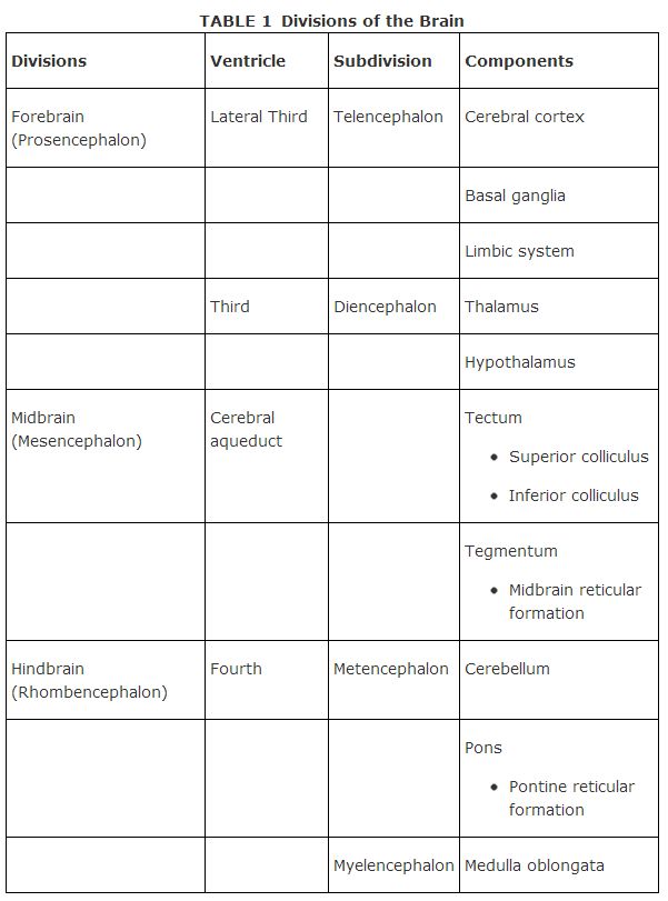
(see below), and the tissue around them becomes the three major brain divisions Table 1
The brain. The three major parts of the brain are
-
the forebrain, the most recently evolved section
-
the midbrain, which contains the upper part of the brain stem
-
the hindbrain, which contains most of the brain stem
The brain has a series of hollow, interconnected chambers called ventricles. The lateral ventricles are in the forebrain and are connected to the third ventricle in the midbrain. The third ventricle is connected by way of the cerebral aqueduct, a long tube, to the fourth ventricle in the hindbrain, which is then connected to the central canal of the spinal cord (Figure ). The ventricular system provides the pathway for cerebrospinal fluid to move in the nervous system.

Figure 1
The Ventricles and the Three Major Parts of the Brain
The forebrain (prosencephalon) consists of two major components: the telencephalon and, below it, the diencephalon.
- The telencephalon (cerebrum) is divided into two left and right symmetrical halves known as cerebral hemispheres. Each hemisphere is divided into four areas called lobes (Figure ), which have different functions.

-
In the frontal lobe are the principal areas that control the movement of muscles.
-
The parietal lobe contains information that regulates so matosensory information (the skin senses of touch, heat, pressure, and pain).
-
The temporal lobe helps integrate sensory information and some auditory information, including language.
-
The occipital lobe (back of the head) is the area from which visual signals are sent.
The central sulcus divides the frontal lobe from the parietal lobe, and the lateral fissure separates the temporal lobe from frontal and parietal lobes (Figure ). The hemispheres are connected by the corpus callosum, the largest commissure (cross‐hemisphere connection) of the brain.
The cerebral hemispheres are covered with a layer of cells called the cerebral cortex and contain the basal ganglia and the limbic system.
-
The cerebral cortex consists of cell bodies, dendrites, the interconnecting axons of neurons, and glial cells (supporting cells). The neurons give the cortex a gray color (hence the name gray matter. Cells connecting to the cortex contain a large concentration of myelin, which is white, and are called white matter.) In humans, the cortex has many convolutions that consist of sulci (small grooves), fissures (large grooves), and gyri (bulges between adjacent sulci or fissures). Most of the cortex is hidden in these grooves.
-
Below the cortex are the basal ganglia, a collection of sub‐cortical nuclei that are involved in movement. (Degeneration of these structures is associated with Parkinson's disease.)
- The limbic system is a collection of numerous brain areas involved in emotion expression. Among the system's structures are the portion of the cortex known as the rhinencephalon, which contains the anterior thalamus, amygdala, septal area, cingulate gyrus, and hippocampus (a structure involved in the processing of memories, particularly short‐term memory). The limbic system also includes neural connections to the hypothalamus.
- The diencephalon, the lower portion of the forebrain, contains the thalamus, and hypothalamus (Figure 3).

Figure 3
Sagittal Section of the Brain
-
The thalamus is a structure through which all sensory information except olfaction (smell) must pass.
-
The hypothalamus is below the thalamus and contains structures that regulate the biological drives (for example, hunger or thirst).
The midbrain (mesencephalon, Figure ) (located between the forebrain and hindbrain) helps regulate sensory processes (such as locating the position of objects in space) and is the location of the dopamine systems involved with performance of voluntary movements. (Damage to these dopaminergic systems may result in Parkinson's disorder.) The midbrain also contains the tectum (which contains the superior and inferior colliculi, primitive centers for vision and hearing) and the tegmentum (which contains the midbrain reticular formation, part of the reticular formation, a structure that runs through both the midbrain and hindbrain and is involved in certain muscle reflexes, pain perception, and breathing).
The hindbrain (rhombencephalon) includes the metencephalon and the myelencephalon.
- The metencephalon contains the cerebellum and the pons (Figure ).
-
The cerebellum is a large structure in the lower back of the brain that coordinates movement and equilibrium.
-
The pons (which means “bridge”) has fibers that connect the brain stem to the cerebellum and also has groups of cells that are important in sleep and arousal, the pontine reticular formation.
- The myelencephalon is below the pons and contains the medulla oblongata, a structure involved with functions such as breathing, swallowing, regulation of heart rate, and other important functions essential to life.
Brain stem is a term used to identify certain brain structures; it consists of the midbrain and parts of the hindbrain (pons and medulla) and connects the spinal cord to the forebrain (Figure ).
The spinal cord. The spinal cord connects the brain to the rest of the body through the peripheral nervous system. The spinal cord is connected to the brain through an opening in the base of the skull and extends to a point just below the waist. It is covered by meninges and is contained within the bones (vertebrae) of the spinal column.